1. Introduction
The heart is a complex organ responsible for pumping oxygen-rich blood throughout the body. Evaluating its structure and function is essential for diagnosing and managing various heart conditions.
Echocardiography, commonly known as an echo test or heart ultrasound, is a non-invasive imaging test that uses sound waves to create detailed pictures of the heart. It helps doctors assess the size, shape, movement, and functioning of the heart chambers, valves, and muscles.
An echocardiogram is one of the most important tests used in cardiology for diagnosing conditions such as heart failure, valve disease, cardiomyopathy, and congenital heart problems.
2. What Is Echocardiography?
Echocardiography is a diagnostic test that uses high-frequency sound waves to produce moving images of the heart.
During the test, a device called a transducer sends sound waves into the chest. These waves bounce back from different parts of the heart and are converted into images displayed on a monitor.
An echocardiogram allows doctors to evaluate:
- Heart size and structure
- Heart muscle movement
- Pumping efficiency
- Heart valve function
- Blood flow through the heart
- Pressure changes inside heart chambers
Unlike X-rays, echocardiography does not use radiation, making it a safe imaging method.
3. Why Is an Echocardiogram Done?
Doctors recommend echocardiography for diagnosing, monitoring, and managing several heart conditions.
3.1 Assessing Heart Function
An echocardiogram measures how effectively the heart pumps blood.
It helps evaluate:
- Ejection fraction (EF)
- Strength of heart muscle contraction
- Overall pumping ability
A reduced ejection fraction may indicate weakened heart function, such as in heart failure.
3.2 Detecting Heart Valve Problems
Heart valves control blood flow between chambers of the heart.
Echocardiography can identify:
- Valve narrowing (stenosis)
- Valve leakage (regurgitation)
- Valve infections
- Structural valve abnormalities
3.3 Diagnosing Heart Failure
An echocardiogram helps determine whether symptoms such as breathlessness and leg swelling are related to reduced heart function.
It can show:
- Weak heart muscle
- Enlarged heart chambers
- Problems with relaxation of the heart
3.4 Evaluating Chest Pain and Breathlessness
Doctors may use echocardiography to investigate symptoms such as:
- Chest discomfort
- Shortness of breath
- Fatigue
- Reduced exercise capacity
3.5 Detecting Heart Muscle Diseases
Echocardiography can help diagnose cardiomyopathies, conditions where the heart muscle becomes:
- Enlarged
- Thickened
- Weakened
3.6 Monitoring Existing Heart Conditions
Patients with known heart disease may require regular echocardiograms to monitor changes and treatment response.
4. Types of Echocardiography
Different types of echocardiograms are used depending on the clinical requirement.
4.1 Transthoracic Echocardiogram (TTE)
This is the most common type of echocardiography.
Procedure:
- The patient lies on an examination table.
- A gel is applied to the chest.
- A transducer is moved across the chest to capture heart images.
It is painless and usually takes 15–60 minutes.
4.2 Transesophageal Echocardiogram (TEE)
A TEE provides detailed images of the heart by placing a specialized probe through the food pipe (oesophagus).
It may be recommended for:
- Heart valve assessment
- Blood clots inside the heart
- Certain structural abnormalities
Local anaesthesia or sedation may be used during the procedure.
4.3 Stress Echocardiogram
A stress echo evaluates how the heart performs during increased workload.
It may involve:
- Exercise on a treadmill or bicycle
- Medicines that stimulate the heart if exercise is not possible
It helps assess:
- Reduced blood supply to the heart
- Exercise-related heart problems
4.4 Doppler Echocardiography
Doppler technology measures the speed and direction of blood flow through the heart.
It helps detect:
- Valve abnormalities
- Abnormal blood flow
- Pressure changes
4.5 Three-Dimensional Echocardiography
3D echocardiography creates detailed three-dimensional images of the heart.
It is useful for:
- Valve assessment
- Treatment planning
- Complex heart conditions
5. What Conditions Can Echocardiography Detect?
An echocardiogram can help identify:
5.1 Heart Failure
It evaluates weakened pumping function and changes in heart structure.
5.2 Valve Disease
It detects problems involving:
- Mitral valve
- Aortic valve
- Tricuspid valve
- Pulmonary valve
5.3 Cardiomyopathy
It identifies abnormal thickening, enlargement, or weakening of heart muscle.
5.4 Congenital Heart Disease
It helps detect structural heart problems present from birth.
5.5 Blood Clots
Certain echocardiograms can identify clots inside heart chambers.
5.6 Fluid Around the Heart
It can detect accumulation of fluid around the heart, known as pericardial effusion.
6. How Is an Echocardiogram Performed?
6.1 Before the Test
Preparation depends on the type of echocardiogram.
For a standard echo:
- No special preparation is usually required.
- Normal eating and medication routines can usually continue.
For certain tests, such as TEE, fasting instructions may be provided.
6.2 During the Test
During a standard echocardiogram:
- The patient lies on the examination table.
- Electrodes may be placed on the chest to monitor heart rhythm.
- A gel is applied to improve sound transmission.
- The technician moves the transducer over different areas of the chest.
- Images of the heart are recorded.
6.3 After the Test
A routine echocardiogram usually requires no recovery time, and patients can return to normal activities immediately.
7. Is Echocardiography Safe?
Yes. Echocardiography is considered very safe because it uses sound waves and does not involve radiation.
It is commonly used in:
- Adults
- Older adults
- Children
- Pregnant women when medically required
8. Understanding Echocardiogram Results
Doctors interpret several measurements from an echocardiogram.
8.1 Ejection Fraction (EF)
Ejection fraction shows the percentage of blood pumped out of the heart with each heartbeat. It helps assess heart pumping function.
8.2 Heart Chamber Size
The test evaluates whether chambers are:
- Normal size
- Enlarged
- Abnormally thickened
8.3 Valve Function
Doctors assess whether valves:
- Open properly
- Leak
- Are narrowed
8.4 Heart Muscle Movement
Abnormal movement may suggest previous heart damage or reduced blood supply.
9. Limitations of Echocardiography
Although echocardiography is highly useful, it may have limitations.
These include:
- Image quality may vary depending on body structure.
- Some areas of the heart may be difficult to visualize.
- Additional tests may be required for complex conditions.
10. When Should You Get an Echocardiogram?
A doctor may recommend an echocardiogram if you have:
- Chest pain
- Breathlessness
- Heart murmurs
- Palpitations
- Leg swelling
- Unexplained fatigue
- Abnormal ECG results
It may also be advised if you have:
- High blood pressure
- Previous heart attack
- Heart valve disease
- Heart failure
- Family history of heart conditions
11. Frequently Asked Questions About Echocardiography
11.1 Is an echocardiogram painful?
No. A standard echocardiogram is painless. Some pressure may be felt when the transducer is moved over the chest.
11.2 How long does an echocardiogram take?
A routine echocardiogram usually takes around 15–60 minutes depending on the type of test.
11.3 Is echocardiography the same as ECG?
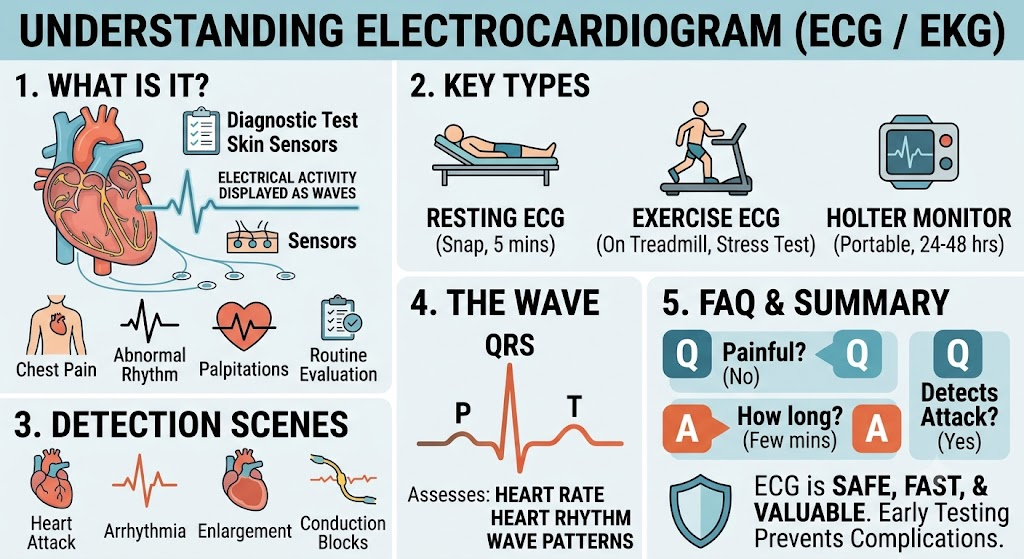
No. An ECG records the electrical activity of the heart, while an echocardiogram creates images of the heart’s structure and function.
11.4 Can an echocardiogram detect heart blockage?
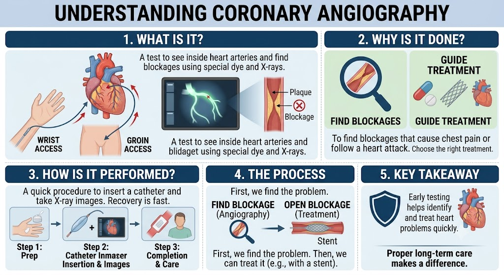
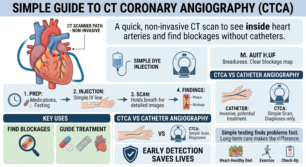
An echocardiogram may show effects of reduced blood supply, but tests such as coronary angiography or CT coronary angiography may be needed to directly assess blocked arteries.
11.5 How often should an echocardiogram be done?
The frequency depends on the heart condition, symptoms, and treatment plan recommended by the doctor.
12. Conclusion
Echocardiography is a safe, accurate, and essential heart imaging test that provides detailed information about heart structure and function. It plays a crucial role in diagnosing heart failure, valve diseases, cardiomyopathy, and many other cardiovascular conditions.
Early evaluation through appropriate cardiac tests can help detect problems at an early stage and guide effective treatment. Maintaining regular heart check-ups, especially for people with cardiovascular risk factors, supports better long-term heart health.